Author:
T. Boland.
History:
3D printing for producing a cellular construct was first introduced in 2003, when Thomas Boland of Clemson University patented the use of inkjet printing for cells.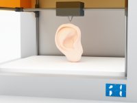 This process utilized a modified spotting system for the deposition of cells into organized 3D matrices placed on a substrate.
Organs that have been successfully printed and implemented in a clinical setting are either flat, such as skin, vascular, such as blood vessels, or hollow, such as the bladder. When artificial organs are prepared for transplantation, they are often produced with the recipient’s own cells.
 More complex organs are undergoing research; these organs include the heart, pancreas, and kidneys. Estimates for when such organs can be introduced as a viable medical treatment vary.
In 2013, the company Organovo produced a human liver using 3D bioprinting, though it is not suitable for transplantation, and has primarily been used as a medium for drug testing.
Example:
Organs that have been successfully printed and implemented in a clinical setting are skin, blood vessels and the bladder.
Description:
3D bioprinting is the process of generating 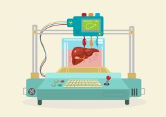 3D printing allows for the
3D bioprinting is being applied to regenerative medicine to address the need for tissues and organs suitable for transplantation. 3D bioprinting has already been used for the generation and transplantation of several tissues, including multilayered skin, bone, vascular grafts, tracheal splints, heart tissue and cartilaginous structures. Other applications include developing
Additions and Criticism:
 Compared with
Publications:
- Mironov, Vladimir, Nuno Reis, and Brian Derby. «Review: bioprinting: a beginning." Tissue engineering 12.4 (2006): 631–634.
-
Derby, Brian. «Bioprinting: inkjet printing proteins and hybrid
cell-containing materials and structures." J. Mater. Chem. 18.47 (2008): 5717–5721. -
Murphy, Sean
v. , and Anthony Atala. «3D bioprinting of tissues and organs." Nature biotechnology 32.8 (2014): 773–785. -
Cui, Xiaofeng, et al. «Direct human cartilage repair using
three-dimensional bioprinting technology." Tissue Engineering Part A 18.11–12 (2012): 1304–1312.